Abstract
Autism disorders are psychiatric abnormalities characterized by improper functioning of the nerves system. It mainly affects the social recognition of oneself including inability to exercise normal executive functions especially those involving learning processes associated with the brain. This paper looks into various developmental syndromes associated with the dysfunction of the nervous system, the relation between head circumference, macrocephaly, and the development of neurological dysfunctions especially in the development of autistic symptoms. The role played by genetic composition and signaling actors in the emergence of autistic disorders will, as well be discussed.
The diversity of the disorder as well as the difficulties associated with the study of the disorder is discovered. The paper also tries to understand the developmental changes in the functioning of nervous system in the developmental stages of a child’s growth. Another area of interest on this paper is to study the genetic influence on the development of autism and other related mental disorders as well as the emergence of the enlarged head circumference before the onset of visible social symptoms of the disorder.
Introduction
The major symptoms and characteristics of Autism spectrum disorder (ASD) are inability to communicate properly, inability to treat others as human beings in relation to social conduct and characteristic repetitive behaviors such as interests in certain activities or objects (Herskowitz, 2000). Statistics on mental disorders shows that ASD has the most inheritable cases of all the other mental disorders with 1 individual out of a population of 150 suffering from Autistic Syndrome Disorder (DSM IV, 1994). Further research showed that boys are more susceptible to the disorder than the girls are with a four times probability to that of the girls (Levitt & Campbell, 2009).
This disorder gets more complicated as its major developments occur at a time when the post-natal stages in early life of a child are occurring. Therefore, as a child’s development process progresses, the condition progresses and gets complicated as well. Thus, the key symptoms of the disorder are always changing as the child continues to develop (Levitt & Campbell, 2009). The disorder may, at times, result to development of other medical conditions.
Medical reports have suggested that the dynamic symptoms of the disorder start appearing very early in a child’s life (Maestro et al., 2002). However, symptoms related to attention and other social cognition behaviors may not be apparent in the first years of life but rather they may show up later but still at early age (Adrien et al., 1992). The major significant and common symptom noticeable to parents is the inability to interact socially and to communicate. Such behaviors are as a result of the improper development of the child’s brain (Herskowitcz, 2000). Much diagnosis has been done so late that there has been no room for much research on the disorder during the early life.
A research conducted by Bartholomeusz and colleagues in 2002, revealed that head circumference in children gives a clear reflection of the brain’s volume while at older ages; the head circumference does not reflect the same. However, the largest percentage of people suffering from autism does not have abnormally large brains and HC. This has prompted researchers to further study the growth of the brain in the early stages of life (Bartholomeusz et al., 2002).
The role played by frontal cortex and the white matter in the enlargement of the brain
To better understand the causes of autism, it is important to study and comprehend the different parts of the human brain (Marlowe, 1992). With the advancement of the imaging devices to facilitate the study of the brain, the examination on this matter has been made much easier. The development of characteristic behavior in autistic people results due to abnormal development of the neuronal system. (Eslinger, 1991)
Studies have identified the abnormal growth of the frontal lobe as one of the most significant cause of autism since they are responsible for the development of major cognitive abilities in children especially during the first years of life when children are constantly growing and developing major features of their bodies. (Courchesne, 2005). At around one and half years, children begin to learn many things including language, self recognition and most importantly, development of reasoning capacities (Herskowitz, 2000). Their ability to relate objects and words is facilitated by the proper maturation and neuronal functioning of the cortices.
The prefrontal cortex plays a major role in the development of inference process in a child. Alteration of the same, consequently, interferes with the proper development of the inference process (Price et al., 1990). Presence of any local lesions in the cortices especially during the early years of life, in that case, interferes with the one’s ability to develop reasoning capabilities (Price et al., 1990) despite the location of the lesion evidenced by patients with lesions at different locations of the brain having similar characteristics such as altered social interaction development and inability to be aware of one’s actions (Grattan & Eslinger, 1991).
Most of the abnormalities of the brain occur in the frontal lobes and they largely affect significant neurons especially the pyramidal cells (Courschesne, 2005). Detailed studies on percellation have shown that abnormalities in Frontal lobes do affect the brain’s ability to function properly affecting the execution of executive functions (Dawson et al., 2002). High head circumferences have been found to relate to abnormal behaviors and development of neuronal system in autistic children (Militerni et al., 2007).
Other studies on neuronal functioning have related abnormal enlargement of the brain to production of high levels of white matter in all lobes especially in the sub cortical part of the frontal lobe which results to abnormalities in the development of other parts of the brain such as amygdala and the hippocampus (Herbert et al., 2003).
Much of the sub cortical white matter appears in the frontal lobe which means that abnormal growth of the cortical connections really does contribute largely to the development of language related disorders (Herbert et al., 2004). Differences in the volumes of cerebral white matter and the cortical may as well indicate lack of connectivity in various regions of the brain, resulting to unintegrated functioning of the rain which is one of the major causes of abnormal behavioral development (Zeiger et al., 2004).
The basic behind the development of autism is a drastic change in the brain size where the brain is extremely reduced at birth but suddenly increases excessively between the second and the fourteenth months of age. (Courchesne & Pierce, 2005). Further studies have confirmed that these abnormal growth patterns of the brain are more apparent in the frontal lobe and other vital areas such as the cerebellum. These characteristics have had long term effects because they cause a slower growth and development of the brain in later years of one’s life (Courchesn & Pierce, 2005).
Further neurological examinations involving diffusion tensor imaging have revealed abnormalities in neocortical circuitry which is characterized by reduced activation of the cingulated circuitry, a vital component of the brain responsible for memory when carrying out various tasks (Luna et al., 2002). A diffusion tensor imaging study carried out by Sundaram and colleagues revealed that the value of the white matter in the prefrontal cortices and in other temporal areas of the brain were different which suggested that there is no integration of the white matter which, then, explains abnormal social behaviors in autistic patients.
Autistic people have also been found to have disrupted white matter pathways as well as disintegration of various regions of the brain (Sundaram et al., 2008). The functional magnetic imaging resonance studies on the brain have revealed that some cortical as well as sub cortical areas of the brain are not well activated due to mutation of the genes responsible for the activation process. The fMRI study of the brain in autistic patients has also revealed reduced connectivity and correlation in functionality of executive tasks such as working memory (reviewed by Minshew et al., 2007).
All these abnormalities of the brain especially those affecting the normal connectivity and integration of all the brain parts indicate that the various parts of the brain do not work together as they should through sending and receiving messages from one part to another which should be facilitated by the various nerve cells (Zikopoulos & Barbas, 2010). Healthy and proper functioning of the brain is enabled by the ability of the brain to convey and integrate messages from the different parts involved in controlling the body activities. The ability of one to reason deeply is facilitated by the integrated functioning of the brain which brings the coherent thoughts and pictures together.
Therefore, perceptions and reasoning thoughts cannot assembled together if the cells of the brain are not able to communicate and work with each other which lead to inability of an individual to process information, most importantly, information involving high order inferences. Alteration of these connections interferes with the brain’s ability to process the basic executive functioning as well. A study by Zikopoulos and Barbas (2010) revealed that the white matter had specifically changed in areas below the ACC (anterior cingulated cortex) and the orbital frontal cortex. These changes resulted to altered interactions between all the prefrontal areas of the cortex which means the lateral prefrontal cortex could easily be affected.
These findings explain why there are disconnections between distant pathways as well as abnormally increased connections between close pathways, both of which affect development of emotion. This lack of adequate connectivity between neurons has also been reported to affect growth of axon which is normally responsible for guidance and development of ability to connect events. These pathway disconnections in conjunction with unnecessarily excessive connections of the brain areas explain why autistic individuals have difficulties changing attention and they behave in a repetitive manner. Treatment processes of autism should, therefore, focus on correcting the signaling pathways because they directly affect the development of events and guidance (Zikopoulos & Barbas, 2010).
One of these crucial pathways is the PI3K pathway which when altered, may cause the brain to process much information in a localized region rather than involving the whole of the brain. Though these alterations may seem small, they all combine and impair the more complex functions of the brain such as communication capabilities. This paper will therefore, look into the possible factors associated with the enlargement of the white matter of the brain. The role played by PI3K which is vital in proper functioning of the brain will be discussed later in detail.
Autism and abnormality of the brain
Courchesne et al (2003) carried out an examination of the brain in an autism diagnosed patient before the symptom become apparent and after the onset of symptoms. The study focused on the analysis of the longitudinal changes observed in the circumference of the head in newly born autistic children and similar examination in children who developed autism later in development. The examination revealed that as the children continued to grow and develop, their head sizes continued to increase as well. The increase was so drastic such that 59% of the examined infants showed enlargements by 2 standard deviations.
This evidenced earlier findings that the enlargement process increases as the affected children develop as well. At approximately two years after the birth of autistic babies, their head circumference had increased drastically (Yang & Gill, 2007). The behavioral and cognitive developments such as self awareness and language development that take place during this period are greatly affected. The development of these symptoms increases as the two hemispheres of the brain enlarge (Herskowitscz, 2000). In addition, the development of major psychological functions is facilitated by the proper functioning and integration between the two hemispheres. The frontal region, the cerebellum and most importantly, the hippocampus, therefore, have to work together in improving the functioning of the brain.
The result of this study confirms the findings of MRI studies which reported that the volume of the brain in autistic children increases up to around 10% above the average volume between the ages of 2-4 years. However, the increase in the volume of the brain may occur earlier and rapidly in severe cases of the disorder. This study focused on the responsibility of the increased head circumference in the phenotyical features of the disorder later in the affected children. The increase in head circumference in autistic infants has been related to abnormalities in cerebral and cerebellar growth later in development (Courschesne, 2003).
Further studies revealed that autistic children encounter maximum enlargement of the brain at 2-4 years of age. This was evidenced during this study carried out using MRI tests which showed the existence of the greatest enlargements on the white and grey matter of the frontal lobes occurring at that age (Carper at al., 2002). Hence accelerated overgrowth of the white and grey matter occurs during the early years of life rather than developing as the autistic children continue growing.
Researchers have also looked into genetic heritability of the autism syndrome disorder as evidenced by the various phenotypic similarities in family members with history of the disorder (Lakshimi, 2007). Although much study shows that disruptions and lack of cohesion in major molecular pathways are the major cause of impaired development of the neuronal system, there are other factors contributing to the same condition but their contributing effects are not adequate enough on their own to cause the development of the disorder. Thus, they only facilitate the continuation of an already existing condition (Nishimura et al, 2007).
Environmental factors ranging from the intrauterine environment to post natal environmental factors (Newschaffer et al., 2007), have been found to facilitate the changes involved in the genetic constitution of an autistic individual which may, consequently, result to further abnormalities in the development of the neurons as well other personality characteristics associated with autism (Pardo, 2005).
Theories on causes of ASD suggests that environmental factors especially those related to routine experiences interact with typical genes resulting to development of patho-physiological conditions which may affect executive functions such as communication abilities and self awareness (Levitt & Campbell, 2009). The initial dysfunctions, thus, provides a basis under which other much complicated conditions can develop.
However, studies on the relation between developmental factors on phenotypic diversity and the genetic contribution to development of ASD have not yet been cleared up although many researchers believe that genetic changes over many generations results to mutations which are then revealed in various phenotypic features (Hammock & Levitt, 2006). Many scientists have done molecular studies on mutated mouse which have produced various models hence providing a basis for studies on etiology of many of the human diseases including autism and related mental disorders (Darnell et al., 2005).
Such studies on ASD and other genetic disorders have, however, been faced with major setbacks as the diagnosis of the disorder focuses more on social interaction abilities rather than neurological examinations (Levitt & Campbell, 2009). This factor has made it difficult for researchers to examine such characteristic symptoms in mice since they cannot have similar social characteristics as human beings do. Furthermore, the single genes available in the mouse mutants hardly provide the complex genetic and phenotypic characterization of the ASDs hence providing inadequate material for the study (Levitt & Campbell, 2009).
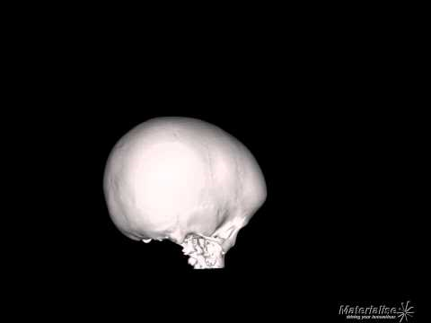
Macrocephaly
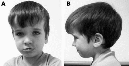
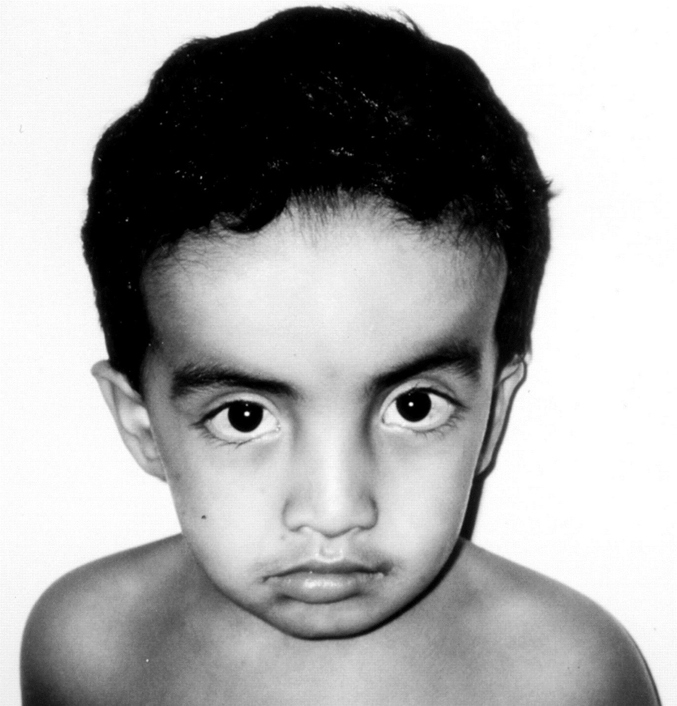
Macrocephaly is the abnormal enlargement of the brain which may occur at or after birth. This condition may appear to be more prevalent than expected with 97% of a 100% population of newborn children having the condition. The frontal lobes in autistic children during their early years of life, probably between two and three years, have the most enlargements evident in the grey and the white matter components of the brain (Herskowitz, 2000).
Mutations of the PTEN gene have been related to development of macrocephaly especially in autistic patients as a significant number of autistic patients have been found to have head circumferences that are between 2.5 and 8 SD more than the normal HC. This finding was found to be equal in both boys and girls (Butler et al., 2005). The development of functional abilities relies on the generation of circuitry which is facilitated by proper neuronal growth as well as adaptive processes resulting from learning experiences (Backman et al., 2004).
Another research, by Herbert and colleagues, sought to understand the physical characteristics of autistic brains in patients with macrocephaly revealed that the brains are not only abnormally large, but have un-proportional sizes on different parts which create an unbalanced condition of the brain. Through the use of IMR technique, clear and detailed images of the brain supported earlier revelation that the white matter component of the brain has the greatest enlargement compared to other parts such as the cortices. As the name suggests, white matter describes the white fatty material which covers nerve cells of the brain to offer protection to the nerve cells based on the insulating properties of fatty acids.
The white matter, therefore, contains many nerve fibers which function to convey information between the different parts of the brain. Further and closer examination on the white matter material was conducted. It showed that the enlargement of all white matter in the various regions of the brain was equal. However, differences in the enlargement rates of the white matter occur depending on the distance through which the white matter material conveys information to and/or from.
In this case, the white matter material, responsible for information transmission between neighboring parts of the brain, seemed to enlarge at a higher rate than those which conveys messages between distant regions of the brain which in most cases maintains its constant volume (Herbert et al., 2004). Generally, major and significant enlargements of the brain had happened in the posterior part of the brain which is responsible for most of the integration functioning of the brain due to its interconnections with the rest of the brain thus promoting the integration of all brain areas for efficient functioning (Schultz & Robins, 2002).
Since the white matter area develops later in life at around two years after birth rather than developing in the uterus, researchers are hoping that postnatal interventions could be availed during this time when the condition has not yet developed completely to prevent the abnormalities associated with the enlargement of the white matter material from occurring (Herbert & Ziegler, 2003). Studies also showed that connections in the brain are more and efficient on individual halves of the brain than they are between the two. This means that information is easily shared and integrated in either of the two sides of the brain than it is between the two halves.
This creates a condition in which each side of the brain deals with its own information by itself without involving the other half, a condition known as lateralization. For instance, the left side of the brain contains much of the information required for language capabilities than the right side (Kemper and Bauman, 1998). Thus, the portions of the brain involved in the processing of language are largest on the left side of the brain, something which may create imbalance of the brain. Therefore, individual functioning and connectivity of the two halves would prevent abnormalities in development. However, detailed studies on the brains of autistic children gave opposite results concerning the sizes of the halves of the brain.
The left side which should be larger than the left appeared to be smaller in these cases hence reversing the lateralization condition of the brain (Herbert, 2005). The areas affected by this asymmetry were the ones responsible for executive functioning as they are responsible for bringing together information from all other regions of the brain. It was also noted that white matter enlargements appeared in children who suffer from Developmental Language Disorder. The similarity between these two disorders led to the conclusion that white matter enlargement is the primary cause to inability to process information in such patients (Herbert, 2005).
Both neuropathological studies and clinical examinations confirm the role played by neuronal dysfunction in alteration of the information processing system of the brain due to poor organization of the dendrites as well as the inadequacy of connectivity between the various pathways, both of which cause changes to the structure of the brain (Minshew & Williams, 2007). These changes present the main causes of altered development of social behavior in young children (Landa et al., 2007).
Conversely, the effects of these genetic changes are facilitated by environmental factors (Cassonova et al., 2002). The development of ASD takes place early in life in the mother’s uterus with major changes occurring in the neuropathology of the brain although the exact timing of these developments has not yet been identified (Luthert et al., 1998). The most evident changes in neurodevelopment of autistic children are on the cerebral cortex and the cerebellum.
The relation between PTEN, macrocephaly and autism
PTEN are mutated genes that cause a condition known as the Cowden syndrome, which is characterized by the development of numerous central growths hence increasing one’s risks to cancer development (Pilarski & Eng, 2004). These mutated genes are inherited and are also found in people with proteus syndrome and another autism related condition known as Bannayan Riley Ruvalcavba Syndrome (BRRS), whose patients have been identified to have similar characteristics to those in autistic people (Zoroglu et al., 2004). The mutations that occur in the PTEN have been identified as the causative factors to the development of nacrocephaly.
Macrocephaly occurs in patients of Cowden syndrome although some may as well be autistic (Gofin, 2001). In addition several studies have shown a close correlation between macrocephaly and the PTEN gene mutation especially in autistic individuals. (Butler et al., 2005). A study carried out by Butler and colleagues involved the introduction of the PTEN mutated gene into 18 patients with co-occurring autism and macrocephaly. After allowing the foreign gene to get integrated in the genetic composition of the patients, examination of the genes showed that 8 out of the 18 patients had heterozygous mutations of the PTEN gene (Butler et al, 2005).
Another recent study on the same revealed that some autistic patients with macrocephaly had missense PTEN genes as a screen on 88 patients with both macrocephaly and ASDs confirmed that one of them had developed missense mutations of the PTEN gene despite the fact that there was no deletion of normal genes (31). This has resulted to routine performance of such tests in patients having any of the three disorders due to several other reports that some autistic patients have the PTEN genes too (19, 99).
However, the symptoms of the three conditions may differ despite their close relation. For instance, it is not yet confirmed if PTEN mutations in autism have the same feature of an enlarged head size (Herman et al., 2007). Similarly, it is not clear if autism itself can result to disruptions of the healthy PTEN gene. It is, thus, important to carry out examinations and find out if the signaling factors that control and regulate the PTEN gene are altered in any way during the development of autism (Buxbaum et al., 2007).
Another study to determine the role played by PI3K pathway in the development of autism was carried using mice. The results revealed that mice which lacked the PI3K pathway had similar characteristics to those in autistic patients, with the most evident characteristic being the inability to distinguish between living things and inanimate objects. This symptom as well as others characteristic to autism has been found in mice that do not have the PI3K pathway which is responsible for regulatory functions of cells (Luis Parada, 2010). Some of its functions are to regulate the size of neurons produced and to control the signaling connections between the neurons.
This characteristic is similar to that found in autistic patients hence the relation. Mutations associated with the PI3K pathway, just like in the case of PTEN, have also been identified in autistic patients. Inactivation of the PTEN genes in the forebrain of a mouse resulted to larger brain development and other behavioral characteristics similar to autistic individuals (Eckhart, 2010). As mentioned earlier, several studies have related autism to the PTEN gene mutations and macrocephaly (Butler et al., 2005). This is evidenced by the presence of PTEN mutations and neuronal related behaviors in Cowden syndrome patients (Butler et al, 2005).
Relation between autism and other disorders
Germline mutations have been used to diagnose clinical disorders such as autism and macrocephly. For instance, in a recent study Butler and colleagues, germline mutations of the PTEN gene were found in three children who had both autism and macrocephaly (Butler et al, 2005). Similar findings were done by another researcher, Heman et al who studied gene composition in children who were not related in any manner those of the above mentioned study but they, too, had autism and macrocephaly. Two of those studied showed presence of mutated forms of the PTEN gene (Herman, 2002).
Due to these findings, it has been suggested that the sequencing of the PTEN gene should be done in all autistic and macrocephalic patients for responsive measures to be taken as early as possible (Butler et al, 2007). This is evidenced by an earlier study which reported a case of a missense mutation of the PTEN gene in one of the 88 autistic and macrocephalic patients although complete gene deletions were not reported. In addition, the phenotypic characteristics associated with PTEN germline mutations may be more than anticipated according to previous studies (Orrio and Galli, 2009).
Deletion of the PTEN gene from the neurons of the cerebral cortex after completion of mitosis in experimental animals has resulted to characteristic poor social behaviors due to neuronal changes as well as progressive macrocephaly (Kwon et al., 2006). The transgenic animals behaved in a manner that is so different from their normal behavior especially on social interaction with other animals. For instance, most wild animals would show much interest in mice but in this case, they hardly interacted with them despite the rare encounter. Instead, the transgenic animals showed much interest on other objects which were inanimate (Moy et al., 2004).
Such changed occurred due to various aspects of the changes experienced in the neuronal regions. Besides enlargement of some parts of the brain, the transgenic animals also showed altered functionality of the axon and dendrites parts of nerve cells which make up the brain (Jarwoski et al., 2005). The mutants also showed overgrowth of fiber tracts as well as increased growth in dendrites and synaptic vesicles.
These results confirmed a previous study which implicated the AKT/mTOR pathway in causing branching of the dendrite system alongside PTEN functioning. In addition, alterations in the development of the neurons results to another disorder called tuberous sclerosis (TS) whose signaling pathways correlate in function to those involved in PTEN mutations. Majority of TS cases have been diagnosed alongside autism as TS patients show autistic behaviors which have been reported to result from alterations in the number and positioning of cortical tubers (Weigel et al., 2010).
However, this report has not yet been confirmed as other researchers have found no relation at all between the location and number of cortical tubers and characteristic autism behaviors (Bolton et al., 2002). A study conducted by Kwon et al in 2006 showed that changes in intracellular processes associated with signaling functions do cause unusual migration of neurons and abnormal extension of neuritis (Weigel et al., 2010). These studies also concluded previous findings that alteration in the PTEN gene, results to loss of organization in the brain whether the alteration involves the complete deletion of the gene or not.
Examination of adult laboratory mice consisting both PTEN positive and PTEN negative species showed that PTEN genes are responsible for not only suppressing tumors, but also regulating the movement of cells within the various brain regions. Surprisingly, the inadequacy of PTEN genes has been reported to be beneficial in that it prevents defective programming of cell death when the cells encounter oxidative stress which can be managed without killing the cells (Li et al., 2003). Studies on PTEN genes have revealed that the gene is a major regulator of the processes involved in brain development (Li et al., 2003). This is the factor which makes the alterations of the PTEN gene responsible for neuronal development involved in ASDs.
Mouse models as research tools
Similar to other scientific studies, research on PTEN mutations was done on mice which appeared to be less interested in other unfamiliar mice. On the other hand, the control mice were very much interested at approaching the strange ones. Exposure of mutant mice to both strange mice and inanimate objects, gave interesting results as the mutant mice had equal interests to the two (Moy et al., 2004). This is the same case with autistic children who show much interest on inanimate things such as toys rather than on people. Additionally, the mutant mice were provided with nesting material but they ignored it while on the other hand, the control mice were delighted and they built up their nest.
The little ones from the mutant mice died more often than those of the normal ones, probably due to inadequate care from their mothers. Through the use of mice, studies have shown that macrocephaly is also characterized by the abnormal enlargement of the hippocampus which resulted to inability of the test mice to interact normally with others. This enlargement was found to be specifically at the dentate region of the hippocampus which explains the strange behavior of the 4 weeks old mice.
A few more weeks later, all mutant mice developed characteristic symptoms. This condition failed to be suppressed by the administration of rapamycin which was given before the onset of symptoms in some mice and after in others. On the other hand, the administration of the same on the non-mutant mice worked very well in suppressing the hippocampus overgrowth and other characteristics associated with the PTEN mutation.
The efficiency of rapamycin was facilitated by the defective apoptotic processes from which it was obtained (Buxbaum et al., 2007). This study, therefore confirmed that PTEN genes are responsible for proper organization of the brain and the regulation of cells’ movement in the brain (Kwon et al., 2006). However, the brain may as well get enlarged due to other body conditions rather than macocephaly and PTEN mutations. For instance, diseases such as Tay-Sachs and Alexander disease may cause the brain to enlarge (Buxbaum et al. 2007).
Research on autism has been faced with some challenges especially due to the diversity of the condition. Researchers should use a wide range of data to make and draw adequate conclusions. For example examination of as many autistic individuals as possible should be made when carrying out these studies (Levitt & Campbell, 2009). Another challenge comes in when there is difficult to find a patient with well defined autism characterized by progressive development of symptoms and showing progressive changes in case of interventions.
These studies should not only focus on the causes of the disease, but also on the other health complications associated with the disorder as well as possible treatment and interventions for the same. In addition, the various characteristics of autism indicate that various parts of the brain are involved in its development and therefore the brain should e studied further (Levitt & Campbell, 2009).
PI3K signaling
PI3K is an enzyme family which is very significant in the body as it regulates major functions of the cells. Some of the most important functions of the enzyme include regulation of cell growth and development. It is also involved in cellular movements hence it is responsible for cancer development. The enzyme is also responsible for a condition known as Long Term Potentiation which is characterized by transmission of signals from two neurons simultaneously (Bliss & Colleridge, 1993). This phenomenon is the reason behind the changing strengths in different chemical junctions (Bliss &Coleridge, 2003).
LTP is a very important enzyme since it plays a major part in controlling learning and memory (Cooke and Bliss 2006). An experiment of this enzyme on rat showed that PI3K inhibitors suppressed the expression of LTP despite the fact that its production is induced [15]. However, the dependence of the expression of LTP on PI3K enzymes has been reported to decrease with time (Blatt, 2005).
PI3K enzyme is also very important in its recruitment of the Mtor protein creating a pathway necessary for phosphorylation of another enzyme, p70S6K, which regulates translational activities during protein synthesis (Bolton et al., 2002). PTEN is equally important as it acts as a gene for suppressing tumors. Loss of its function due mutation or deletion of base pairs results o unusual growth or death of cells. When PTEN mutates after mitosis, abnormal enlargement of cells results to formation of differential ganglion cells which further results to a condition, Lhermitte-Duclos which is closely related to Cowden syndrome (Kwon et al., 2001).
Another possible cause of autism as well as other disorders such as schizophrenia is the activation of the immune response during a time which the brain is developing (Levitt & Campbell, 2009). Hence the link between mutation inheritance and the development of altered brain functions which then result to abnormal behavior in PTEN and autistic patients. PTEN genes are also involved in the repression activities of P13K pathway and have also been linked to implications of ASD (Campbell, 2006). Altered functioning of the PTEN gene has also been associated with other mental disorders such as schizophrenia (Hippisley-Cox, 2007).
Interestingly, families with history of schizophrenia are at lesser risks of getting cancer compared to the rest of the population (Licterman, 2004). This confirms that certain genes that result to mental disorders may be beneficial in reducing risks of cancer in those patients (Serajee, 2003). Environmental factors have also been found to later the phenotypical outcome of autism by affecting the regulatory mechanism of the PTEN-PI3K pathway which directly affects neuronal development (Schanen, 2007).
Conclusion
Studies indicate that the major causes of autism are genetically related although substantial evidence on environmental causes has also been provided (Levitt et al., 2009). Different individual suffering from this disorder have been displayed to have diverse symptoms and characteristics as well different severity of the disorder. In the efforts to understand the phenotypical characteristics associated with autism, researchers have discovered that the enlargement of the brain in autistic patients is related to another mental condition, macrocephaly. In fact, the enlargement of the brain is the only feature that seemed to be common to all autistic patients.
The cause of this overgrowth has been invoked to be the inability of the nerve cells to integrate the functioning of the synapses in the brain. PTEN which is a gene involved in cancer development, has also been identified to play a major role in the development of the neuronal system as well as a tumor suppressor (Meahama, 2007). The dysfunction of the synapses which starts at childhood continues to adulthood although its effects are largely determined by one’s experiences and other environmental factors (Cook & Bliss, 2006).
These factors continue to contribute to the remodeling process of the nervous system and increase the transmission of synapses. These developmental processes have the possibilities of altering the development of the disorder making it only recognizable later in life rather than in the early years (Herskowitz, 2000).
Due to the changes involved in the brain as a result of aging such as memory loss, scientists should take much more effective measures to prevent misdiagnosis of this disorder at later years of life (Cassanova, 2003). Further studies should also be done to determine the exact timing of onset of features associated with autism as well as the developmental course of the disorder.
Studies on cellular and neuronal systems in autistic patients have led researchers into concluding that changes in these systems are the reasons behind the unusual behaviors evident in autistic individuals (Kwon et al., 2006). Studies have also shown the relation between mutations in the PTEN gene and the PI3K pathway which should, under normal circumstance, facilitate neurodevelopment (Moy et al., 2004). Research on the inhibition of PI3K pathway could be of much help in developing pharmaceutical interventions of this disorder. In addition, with the discovery of the parts of the brain involved in these abnormalities, detailed examination on the same regions can now be done to develop treatment strategies specific to those areas (Herbert et al., 2006).