Abstract
The use of Electroencephalography (EEG) has become a core factor in determining the levels of aspect for growth and development in the management of mental health Over the years. Electroencephalography has been used by many health practitioners in the diagnosis of diverse brain conditions and ailments such as epilepsy, stroke, and tumours among others. In the past, the problem of detecting various mental ailments and conditions was prevalent. This was blamed for lack of effective and efficient devices for detecting and diagnosing such diseases. In addition, physicians and medical practitioners were unable to detect abnormalities in the brain waves due to lack of a device that would be able to carry out diagnosis of such diseases. It is without doubt that the development of Electroencephalography (EEG) among other related devices has played a critical role in the management of mental conditions.
The development of Electroencephalography is a major step towards the use of basic signal processing, EEG recording and data analysis, a factor that brought a greater reliance on the device. The first chapter one brings out the background and depth of the study. In the second chapter, a further understanding of the device has been generated and also an overview of Electroencephalography (EEG) in terms of data analysis, recording and basic signal processing has been explored. Chapter three offers a review of EEG, and this comprises evaluation of Electroencephalography (EEG) in relation to other interfaces. The use of various literature materials has also been employed to conduct a thorough review of the relevance of improving the effectiveness and use of Electroencephalography. A thorough assessment has equally been conducted on similar systems with various comparative and critical commentaries.
Besides, the chapter provides a critical analysis of the devise, its benefits in mental health diagnosis as well as the drawbacks of using an Electroencephalography device with brain computer interface (BCI) and other imaging approaches. An in-depth understanding of the role of Electroencephalography (EEG) in mental health diagnosis as presented in chapter three aids in preparing the reader for the results expected in chapter four and five. The study on chapter three provides a firm background that employs primary data to generate results for analysis and discussion in chapter four and five.
Chapter four takes a comprehensive analysis of data and reveals the magnitude of the importance of Electroencephalography (EEG) in the medical field. Finally, the conclusions and recommendations section in chapter five calls for further research and intrinsic focus by institutions of higher learning in conjunction with their administrations to design more effective systems of dealing with mental complications.
Preamble
An understanding of the issues involved in any research is important to developing a clear picture of the significance of the research and also plays a role in ensuring that the research approaches employed are placed in their right context. This chapter presents the methodology, significance and nature of the research with the aim of developing a clear picture of the need for research, how the research will be undertaken and the significance of the study on existing knowledge and practice within the business community and society as a whole.
The Electroencephalography (EEG) which is also known as a brain wave test, is a very important examination that has been developed in the recent years. Isley, Edmonds and Stecker (2009, p. 370) observe that the test has been very critical in the medical field since it has aided healthcare providers in the management of diverse brain conditions such as epilepsy, stroke, and tumours. Relevant studies posit that until recently, medical practitioners were unable to detect mental conditions, a challenge that many analysts attribute to lack of a device that may be able to detect abnormalities in the brain waves (Claus et al 2000, p. 81).
Background
From an in-depth review of literature, Allen (1997, p. 239) points out that EEG was developed in the late 18th century. Bayliss (2000, p. 188) exposes that the technology was used to record and measure brain activities of animals such as monkeys and rabbits in Liverpool. Numerous psychologists have utilized this technology in different parts of the world and have been proved to be effective in monitoring brain function. For example, Vladimir Neminsky (a Russian psychologist) used EEG in 1912 to analyse the potential of a mammalian dog’s brain (Allen 1997, p. 239). In the year 1914, this psychologist realized that EEG had the tendency of causing seizures in the brains of animals. In addition, Hans Berger (a German scholar) used EEG device on human brains in 1924. He expanded his research which eventually led to the rise of the name of the device known as electroencephalogram (Bayliss 2000, p. 188).
The development of EEG was later confirmed by other scholars and regarded as a major development in the medical history. In the mid 19th century, numerous scholars explored and did further research on the improvement of EEG in order to reduce the risks of seizures (Kostov 2000, p. 207). Still on the same epoch, further studies confirmed that despite the improvement, the device increased the prevalence of seizures among epileptic patients. In 1950s, advanced developments were made on EEG in order to make it possible to map electrical activities in the brain surface even for patients in remote areas (Bayliss 2000, p. 188). For this case, the wireless device was developed and which made it easier to monitor brain functions of patients without restricting them in ambulatory units. This made it possible for patients to interact with the public and lead their normal life while the monitoring test could even be prolonged for months. Prior to the development of EEG, numerous improvements on design had been made making it convenient to perform large-scale activities in neuroscience (Kostov 2000, p. 207).
Under normal circumstances, the human brain is expected to respond to treatment since everything is related to emotions and signals that are transferred to the brain through the nervous system (Smith et al (2001, p. 954). A quantitative study is therefore necessary in order to determine how the brain functions bearing in mind that emotion alone cannot be used to assess the working of the brain.. it is against this backdrop that over 60% of developments in neuroscience have made it possible to understand human brain codes through a computer model. The latter has also been instrumental towards the determination and prediction of brain activity patterns.
The field of mental health has significantly benefited from Electroencephalograph (Jose et al 2002, p. 113). It is worth noting that Electroencephalography devices that have been developed aid physicians in recording spontaneous electrical such as diagnosis of epilepsy, studying disorders that are related to sleep, assessing the brain nature of individuals who have been admitted while in comma and also assessing any form of loss in consciousness that may be encountered by a patient with mental problems. According to Grønning et al (2005, p. 400), the use of Electroencephalography has been key in diverse clinical circumstances some of which include determination of proper wean anti-epileptic medications, prognostication of coma patients, conducting adjunction tests related to brain death and differentiating catatonia and other related psychiatric syndromes from organic encephalopathy.
It is important for the patient who is preparing to undertake EEG test to inform the doctor when he/she is in any kind of medication. For instance, tranquilizers and certain sedatives, sleeping aids as well as drugs used to relax the muscles should not be taken before the EEG examination is executed to a patient. The aforementioned drugs are known to interfere with test results since they can easily modify the electrical activity of the brain. In addition, foods that are rich in caffeine should not be consumed at least twelve hours prior to the EEG examination. Finally, the hair surface should be made as clean as possible bearing in mind that the scalp holds the electrodes during the test.
According to Bayliss (2000), the Electroencephalography (EEG) test helps to measure and record vital electrical activities in the brain. EEG has special sensors called electrodes that are place on a patient’s head and then connected to a computer through some wires. In the meantime, the sensors enable the computer to record brain activities and also examine certain conditions. Allen (1997) elucidates that this test can be used to locate seizures, abnormal conditions and impairments in the brains. Notable ailments that can be detected using this test include stroke and epilepsy.
It is also important to mention that there are myriads of reasons why EEG test is done. Moreover, there are specific procedures that are used to conduct the test. In-depth analysis of literature has also revealed that EEG test requires special preparation and that there are diverse risks associated with it. Furthermore, there are numerous issues facing the use of EEG devices for patients due to their designs. It is against this backdrop that there is need to address the issue of design improvement of a wearable EEG device.
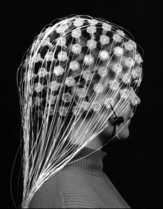
As illustrated in the above diagram, Electroencephalography (EEG) uses multiple electrodes that are placed on strategic points on the scalp of the patient in a bid to recording the waves that are created from the electrical activities taking place in the neurons of the brain. The recording of the brain waves usually takes very short periods of time that may last from 20 to 40 minutes and from the information collected, the medical analysts may be able to determine the disorder that the patient is suffering from (Chua et al 2009, p. 485).
There are myriads of past empirical studies which have demonstrated that a functioning magnetic resonance imaging (fMRI) can be made use of in detecting activated brain areas as a result of the images which are produced (DuanYu et al 2010, p. 110; Liamsuwan et al 2000, p. 27; Knott et al 2001, p. 110). Even so, the emergence of Electroencephalography (EEG) has been very useful in measuring the fluctuations in voltage that occur in the neurons brain due to flows in ionic current which creates a wave that is used for communication among various pathways of the brain. Kleiter et al (2010, p. 750) highlight that the devise is able to find and record electrical activities that take place in the neurons of the brain and also transmits this information in form of waves that are eventually recorded on paper.
It is imperative to highlight that the development of Electroencephalography (EEG) has been regarded as a major milestone in the medical industry. According to Hur et al (2010, p. 186), this invention can be seen in the progress made by doctors and surgeons in monitoring the activities in the brain during medical procedures. The current medical practice has embraced wide use of Electroencephalography (EEG) and has been very critical in neurological applications especially in epileptic cases.
Additionally, besides being used as a first line method in diagnosing focal bran disorders, stroke and tumours, Electroencephalography has also been used to monitor patient’s condition while under the effect of anaesthesia. The latter is a common drug that is given to patients during surgeries. Lee et al (2010, p. 246) reiterate the same in their publication that Electroencephalography has continued to be a valuable tool which bedsides having a limited spatial resolution, has been widely embraced and utilized by physicians to determine brain status. This is due to the fact that it has been designed in such a manner that it can detect the brain waves necessary for the diagnosis and treatment of mental problems.
The paper will explore the background information with regards to the use of the Electroencephalography (EEG) device in the diagnosis of the correct type of mental disorder that a patient may be suffering from. In addition, further research will indeed be conducted to find out how research analysts and other stakeholders in the medical field are working extra hard to try and find out how to improve on the design of the Electroencephalography (EEG) device so as to increase the reliability of the results that are recorded in form of waves. On the other hand, this chapter will try and find out several improvements that have already been made on Electroencephalography (EEG) in a bid to increase the reliability of the results that are recorded. This chapter will also go further and explain in details how the Electroencephalography (EEG) works and the procedure that is to be followed during the use of the Electroencephalography (EEG) in the diagnosis of mental disorders.
Problem Definition
The application of Electroencephalography (EEG) as a means of detecting mental conditions and determining the best diagnostic methods has been intensified globally over the last decade owing to the shifting systems of consumerism that have curved a new niche for healthcare providers in the health industry. Mental healthcare providers have therefore proliferated and sought to conterminously apply the use of Electroencephalography (EEG) in the diagnosis and eventual treatment of various medical conditions. It is worth noting that a close correlation seems to be unmistakable between the applications of Electroencephalography (EEG) with the affect of improvement of mental heath condition detection. Perhaps, this appears to be based on the overall emergence of the practice of enhancing efficiency as medical practitioners in diverse health centers assimilate the new mode of using the EEG.
There is no doubt on the importance of the role played by Electroencephalography in conventional cognitive neuroscience and clinical diagnosis. For instance, recordings from an Electroencephalography (EEG) device especially for patients with epilepsy have been used for predictions of seizures. Besides, extensions from neuro-feedback have been considered to be very crucial for brain computer interfaces (Sipilä et al 2000, p. 203). It is apparent the device plays an important role in developing the confidence that healthcare providers have in diagnosing mental ailments and is equally instrumental in ensuring that there is a fast and efficient detection and diagnosis of mental conditions. The role played by Electroencephalography (EEG) in the current healthcare environment makes it an important operational variable.
In the past, the diagnosis of mental disorders was a little bit cumbersome due to lack of vital machines that would be capable of transmitting information from the neurons in the brain so that a proper study and correct diagnosis could be carried out. The medical analysts used large MRI machines that were quite bulky and expensive for any given hospital to afford and as such, few patients were able to access the use of the machine to get the correct diagnosis.
This prompted research analysts and other major stakeholders in the medical industry to come up with a device that would be able to measure any change in feelings experienced by patients before or during the testing period. That is when research analysts were able to develop the Electroencephalography (EEG) device amid great controversy of the disadvantages in terms of inflicting fear in the patients during the testing period.
The purpose of this as study is to research the design improvement of a wearable EEG. Having enhanced EEG tools has become an ideal way for healthcare providers to enhance the effectiveness in conducting measurements of brain waves and detection of possible mental health conditions. The study aims at providing results that will aid in the development of a correct view of adopting efficient measuring methods and therefore create the necessary supportive frameworks that will reflect improving EEG design and higher quality
The preliminary or background survey reveals that though tremendous development has been attained in addressing ways of enhancing the designs, practical implementation of the roles is far from attaining efficiency that researchers seek. Dynamics that are definitive of technology and practical factors that have to be considered in the business environment are some of the factors that appear to affect businesses in implementing development plans. It is thus evident that there is need for a critical review of enhancing EEG designs to determine both the practical and theoretical systems that have been put in place and what should be done to address the areas that are lacking.
It is important to mention that some of the notable philosophies that guided this research include positivism, interpretivism and realism. Through those philosophies the researcher was able to consider all the possible situations and factors to consider during the improvement of the design for Electroencephalography (EEG) device. These philosophies made it easy to analyse the influences, effects and benefits associated with the use of Electroencephalography (EEG) devices. Moreover, they made it possible to examine the numerous adjustments that need to be made on EEG devices in order to enhance better diagnosis of the mental disorders among patients. Pointless to say, applying the philosophy of realism helps one to have a perspective toward helping both patients and medical professionals.
Despite the challenges and barriers to implement use of EEG technology in hospitals, the benefits can not be quantified. Information derived from previous results has shown that EEG device has gained public acceptance. Therefore, one has to be optimistic that serious improvement tactics will eventually result to numerous benefits where no index can measure. Using the interpretivism philosophy, opinions made from the research were analysed to make suggestion on how to improve the wearable EEG device. This was an added advantage to make all the abstract opinions into reality, a factor that will help to boost the reliability and performance of EEG tests. In a nutshell, the philosophies aided in addressing the general problems both in the social and environmental sphere making it possible to draw conclusion on how to modify the EEG designs.
Literature Review
Introduction
This chapter forms the major section of this study as it provides a link and a holistic overview of the external outlook and implications of all related facets. In this case the chapter inculcates the relevant information that seeks to ensure strong support for the different findings of the subsequent chapters. To effectively anchor the feasibility of the thesis statement, this chapter explores the available literature on how much improving the design of an Electroencephalography (EEG) can be created by implementing technological strategies in coming up with advanced tools. Besides, through exploration of the different theoretical perspectives, the chapter evaluates varying perspectives on the importance of having an improved EEG tool. It also gives a holistic criticism by analyzing the challenges and strengths of improving the designs of the Electroencephalography (EEG).
Type of electroencephalography (EEG)
Schaefer, Vlek and Desain (2011, p. 100) indicate that the Electroencephalography (EEG) records the brain waves in three main ways and that the brain waves that are present in the results are different since the wave lengths differ from each type of recording to the other. The first is the wired Electroencephalography (EEG) which is a bunch of wires that are connected from the Electroencephalography (EEG) device and linked to the scalp of the patient through the use of multiple electrodes. Besides, Greensite and Huiskamp (2000, p. 1253) further highlight that the electrodes that are connected to the scalp of the patient will then transmit the information to the Electroencephalography (EEG) device that will then translate this information and record it in terms of waves.
The second type of Electroencephalography (EEG) wave recording is by the use of a wireless Electroencephalography (EEG) device. Burghaus et al (2007, p. 45) point out that the main difference between a wired and the wireless Electroencephalography (EEG) device is that is the wired, the Electroencephalography (EEG) device uses wired that are attached through multiple electrodes to the scalp of the patient while in the wireless Electroencephalography (EEG) device, the brain waves are transmitted without the use of wires to connect to the patient’s scalp. In the wireless Electroencephalography (EEG) wave transmission, doctors and specialists insert wireless Electroencephalography (EEG) sensors into the patient’s brain which are then going to be used in the transmission of the waves to the Electroencephalography (EEG) device that will then translate the information onto wave patterns. Research analysts have found this type of transmission to be more effective than the use of wired transmission because the waves that are recorded in this type of transmission are more accurate than those that are recorded from the use of the wired transmission.
The third and last type of Electroencephalography (EEG) is the filtered wireless wave transmission. Stavtsev and Ushakov (2010, p. 261) indicate in their publication that the main difference between the wireless Electroencephalography (EEG) transmission and the filtered wireless Electroencephalography (EEG) transmission is that in the wireless Electroencephalography (EEG), the wireless sensors are inserted into the brain matter so as to be able to collect more accurate information while in the filtered wireless Electroencephalography (EEG) transmission, the wireless sensors are attached to the patient’s scalp in a bid to collect the necessary information that is to be transmitted to the Electroencephalography (EEG) device.
In the filtered wireless Electroencephalography (EEG) transmissions, the wireless sensors which are placed onto the patients scalp record the same data as the wired Electroencephalography (EEG) transmission. However, the main difference between the two is that, in the wired Electroencephalography (EEG) transmissions, the information is transmitted to the Electroencephalography (EEG) device through wires that are connected to the electrodes while in the filtered wireless Electroencephalography (EEG) transmissions, the collected information is transmitted to the Electroencephalography (EEG) device through wireless means and then recorded in terms of waves (See appendix I).
Signal Processing and recording using an EEG
Flexer (2000, p. 396) indicates in his publication that an EEG works through macro electrodes and microelectrodes among other components to conduct its various factions. A macroelectrode, whose measurement in terms of size is 5 and 10 mm wide can be described as a cone-shaped metal disk made silver chloride, silver, gold and tin. Wu et al (2012, p. 354) indicate that the popularly used macro-electrodes ar5e normally meant for scalps and usually work for a limited time or session. These can be placed within an individual’s scalps or affixed on top it. While they are non-toxic and safe, an enhanced EEG can be designed to be used safely for longer periods. The macroelectrodes record data from an individual’s brain and the data are referred to as ECoGs. Improving the design of the EEG will be critical in enhancing its measuring activity. Väisänen et al (2008, p. 101) cite that this is due to the fact that the measurements at the electrodes happen at the neutral reference and active sites.
The process of recording signals requires preparing patients for the tests. This according to Howland (2006, p. 12) is done so as to obtain relevant data and clean signal. This process calls for abrading each electrode to remove the dead skin cells which hamper with proper conduction of electricity. Besides, the cleaning reduces galvanic skin response (GSR) or the electrodermal activity. Sarkar, Barks and Donn (2008, p. 120) indicate that an improved EEG device is critical in changing the whole process of prepping. This is because in the imaging of the brain, an improved EEG makes the process easier and faster by employing new sensor technologies.
Resource and Requirements
In order to accomplish the objectives in this research study, there are myriads of requirements that were employed. Moreover, there are vital resources that were gathered before the actual research activity commences. It is important to mention that resources and requirements will highly depend on the method to be used during research. For instance, one will require tools for experiment such as EEG devices. Moreover, other resources include writing materials such as note books and pen for writing sown information obtained. Depending on the methods to be used in collecting data, one might need to have tape recorders and a video camera to record details that can not be put down on a paper. Time is also a vital resource and that it would be adequate to enable the researcher to cover up all the intended activities effectively. Basically, such a study can not be carried out without the help from the respondents. In this case, one should have interview questions, structured questionnaires and other assistive devices in case of experiments.
One of the major requiems is that all the viral resources should be made available before one commence the study. The other requirement is that ethical considerations should be adhered to during the research process. That notwithstanding, it is recommended that the researcher have a research guide in order to align with the research context. Since most of data to be used will be derived from secondary sources, the researcher will be required to collect numerous books, journals or publication to use while reviewing the literature of past research. This will help in identifying the impending research gap and appreciate what has already been done previously (Allen 1997, p. 238). Moreover, the researcher is required to conduct research that is science-oriented. In this case, scientific procedures will be followed to enable one to test the research hypothesis and also overcome bias. In order to execute a concrete and comprehensive research on how to improve designs for wearable EEG devices, there are numerous aspects that were considered. These included psychological, historical, theoretical and institutional background of the study which will be discussed below.
Psychological Aspects
Recent survey results have revealed that there are diverse psychological aspects that influence the function of human brains. Moreover, emotional state of a patient highly determines the reliability, accuracy and effectiveness of the results obtained from EEG test (Feige 1996, p.253). Just like it was mentioned in chapter two, studies have shown that fear affects the accuracy of results obtained using the brain test devices. Evidence derived from surveys revealed that most patients get nervous when they get onto contact with the multiple electrodes on the scalp (Birch 2000, p. 193). For this reason, studies have shown that it is important to ensure that patients’ emotional and environmental status is stable before the test is done. In-depth analysis has illustrated that anaesthesia helps to relax the patients mind from fear and anxiety. For this reason, medical experts inject drugs on patients during the test and this has been proved to cause adverse effects to the results. From a careful review of literature, Luck (1990, p. 613) notes that EEG should be designed in a manner that they are able to detect transmission of waves from the brain regardless of the psychological state of the patient. Moreover, they can be designed in a way to cause anaesthesia to the patient, a factor that will elevate the degree of reliability of the test results (Makeig 1995, p 214).
Theoretical Background
According to Matt (1998, p. 853), EEG has the most advanced form of brain computer interface that has highly made an impact in neuroimaging. Further research has illustrated that there is need for advancing the technology to make the test more convenient. Scholarly studies have shown that the tests do not cause any side effect or pain to the patients. Notably, during the test, doctors attack more than 16-21 electrodes on ones scalp. Of worth to note is that these electrodes can scare children who need to undergo the test. For this case, Pfurtsheller (2002, p.48) advocates that use of wireless transmission device be used to avoid causing anxiety to patients. Theoretical studies have revealed that some EEG devices trigger development of brain seizures especially for individuals suffering from epilepsy. This brings one to the pint that designs for EEG devices should be made convenient in order to ensure that the test procedures do not interfere with the patients’ anatomy (Allen 1997, p. 239).
Studies have revealed that some of the wearable EEG devices are very bulky and expensive making it hard for hospitals to purchase them. Moreover, most of the medical experts in ambulatory units opt to forego those that are bulky and prefer those that are more portable. Once of the perceived benefit of implementing portable EEG devices are that they enable to offer services to clients in case of emergences.
Although the introduction of this device in the medical field elicited heated debate among the major stakeholders and other research analysts, the medical analysts went on ahead with the use of the Electroencephalography (EEG) in the diagnosis of the mental disorders that were considered to be one of the diseases that were the most difficult to diagnose.
However, limited research exists on the effectiveness of the Electroencephalography device especially due to its limited spatial resolution. Though many researchers have concluded that an EEG is vital to the effectiveness of detection of mental problems, some have failed to describe or define the problem of an EEG in such a manner that reflects its importance. This can be due to the fact that they perceive the EEG to be limited in its functions and laud the emergence of technology as a means of developing new modes.
An understanding of the purpose of this study is very critical since it links the study on the design improvement of Electroencephalography (EEG) to its expected impacts on the diagnosis of mental related conditions. Due to the advancement in technology and which is increasingly being felt in all sectors, its affects in the health sector is particularly crucial. This study seeks to establish the effectiveness of developing and designing better Electroencephalography with particular focus on its ability to effectively detect diverse rhythmic and transient activities of the brain, the research results will provide timely information that will assist medical experts in ensuring its proper application. Notably, the use of Electroencephalography (EEG) has not been intensively studied. As such, this particular study will be critical in developing policies and standards for its applications.
From the study results based on the views of improving the design of wearable EEG, it will be possible to make particularistic conclusions on its efficacy in terms of detection of patients’ mental conditions. Technologically, this study will be of great essence in seeking harmony between the applied EEG methods of detections and the existing changes and demands in the marketplace. From the results, it will be possible to explore better design of wearable EEG tools that can be able to further enhance advanced interactions between patients and health gadgets.
An understanding of the correct theoretical consideration behind every study is very crucial in gathering an intrinsic evaluative capacity for the process being used and the later analysis of the results. The level of competition in the global market arena couple by the demand for effectiveness in the provision of mental health services have evoked the need for heath care providers to adopt technology and better product designs. As such, new methods and strategies being assimilated require strong local derivation and high level appeal to gain the required standards of operations.
According to Antonenko et al (2010, p. 425), Electroencephalography (EEG) and related technologies have become important tools in healthcare provision due to their unique and resourceful features such as smart structures, event driven messages and notifications. These features play a crucial role of aiding caregivers to check for vital signs and obtaining patients’ information necessary for effective treatment, offering personal healthcare and assisted living. In mental health monitoring, the application of Electroencephalography (EEG) requires measuring a patient’s signals or important symptoms. The latter are used by physicians to make diagnosis and are commonly termed as vital signs. In agreement, Barnes, George and Ng (2008, p. 905) demonstrate that vital signs in a patient vary and may include acceleration of body core temperature, saturation of blood pressure and oxygen (SpO2), electrocardiogram (ECG) and heartbeat rate depending on a health problem. It is imperative to point out that while vital signs are critical for making effective treatment, they may not be sufficient enough as reasons for making a comprehensive diagnosis. As such, medical professional need context information to make valuable diagnosis.
Weinstein, Zhukov and Johnson (2000, p. 1059) point out that in monitoring healthcare provision, context becomes an important aspect which encompasses an entity and characterizes its position and situation. Electroencephalography (EEG) employs the use of context to link an application with its user thereby building an interaction. It is important for the sake of understanding to highlight at this point that contexts vary in types and the primary ones include activity, time, location and identity. Hung et al (2005, p. 1053) point out that context can be high level with user activities which are complex, low level like bandwidth, temperature and time, and can consist of information that is either explicit or implicit. In a healthcare setting, context is an important component which doctors and nurses to characterize fully a patient’s health status. This may be in terms of age, stress levels, location and low or high body temperatures among others. Mental health monitoring therefore plays a crucial role in deriving informational items from patients such as a patient’s specific condition, medical knowledge, and current vital signs as well as medical history. This information as Hung et al (2005, p. 1053) and Wang et al (2008, p. 916) continue to posit aid in reducing medical errors and false positive incidences.
Recent studies have shown that a wearable EEG device is very convenient and has numerous uses. Nevertheless, the device requires constant monitoring through use of inbuilt ambulatory services (Wolpaw 1997, p.529). Since this test is non-invasive, the patients can go on with their daily activities while recording through cameras takes place. However, to make it more conveniently, it is advisable that patients undergoing EEG monitoring to take their time off to allow appropriate test without being disturbed. Recent survey report revealed that once the EEG monitoring device was initially developed, it was very bulky (Vaughan 1996, p. 425).
Currently, it is convenient to conduct a research and establish the ways in which the wearable EEG device can be improved to enhance portability. It is definite that patients would often get tired of being position in an ambulatory unit where they feel isolated (Roberts 2000, p.56). For this reason, there is need to envision the wearable EEG device such that it become small and convenient to be used by patients even in public places. Moreover, making the design smaller will help medical specialists to use it for months, days or weeks continuously to monitor patients’ brain activities (Wolpaw 1997, p.530). In line with this, Costa (1998, p. 105) writes that improving the design of EEG device will help medical specialists to prolong the monitoring period for chronic conditions, a factor that will greatly increase the end-user credibility of the test. For this reason, this research emphasize on how improvements can be made on EEG devise to make its design convenient for use among psychiatric patients.
An understanding of the theoretical aspect and the nature of the environment are just as important as determining the internal organizational systems in developing effective operational strategies. An understanding of the need to improve wearable EEG is therefore incomplete if an understanding of the operational environment and the factors that may affect it is not developed. The primary research questions for this study will be as follows:
- What is the significance of improving the design of a wearable Electroencephalography (EEG)?
- What are some of the environmental consideration and patient interactions with the EEG that needs to be improved?
The following hypothesis has been developed to aid in addressing the research questions.
- Improving the design of an Electroencephalography (EEG) is critical in enhancing its ability to effectively detect diverse mental conditions.
- There is a relationship between improvement of an EREG design and efficient measurements of wave patterns.
Significance
The study is founded on the important role an advanced EEG plays in effective detection and relation functions in mental health. No one can deny the role played by globalization in affecting the dynamics in healthcare operations as it structures how a healthcare provider defines the requirements of its operations. The application of EEG as a means of setting a stage for diagnosis of mental conditions is a platform upon which a healthcare can be aligned with the effects of globalization and gain from advances that are being made in mental healthcare. The role played by an effective EEG in measuring rhythmic activities and transients makes improving its design an important step that health organizations can use to enhance their service provision and sustain their operations.
This study targets to restrict itself on the background information and psychological aspects related to EEG device. This implies that one has to review the literature to understand in-depth facts about the emergence, applications, challenges and significance of EEG device. This will be done by giving a theoretical, historical and institutional background of EEG test. The study will also address how improvement of EEG design will enable medical experts to examine problems associated with brain function such as tumours, coma and memory impairments. This will be achieved by reviewing the literature in order to understand the evolution of diverse types and designs of EEG device and how current models cabn be improved. In this context, the research will also examine how, medical experts and patients will benefit from the suggested designs.
It is thus apparent that the study whose main aim is to find out how to improve on the design of the Electroencephalography (EEG) device and how it will be critical in increasing the reliability of the results that are recorded in form of waves. Performance development especially in the health sector have over the years shown close correlation to the level of growth that are displayed in the quality of tools used in measuring and/or provision of healthcare. The existence of gaps presented by the practice of detecting various health abnormalities is evidenced by the occurrence of issues related to out-dated machines, fast-paced changes in technology and cost. There is need to set in place systems which will aid healthcare providers in developing better health provision platforms. The existence of gaps needs not to be the case considering the role played by research in adding to development that is being made in practice. Such an understanding not only aids healthcare providers in developing effective strategies that may not suffer the failure of previous practices but also aids future researchers in addressing areas that are important. There is no doubt that the dissertation is important in ensuring that developments are made in enhancing the designs of EEG’s.
Effectiveness of an improved Electroencephalography
Electroencephalography like other tools designed to measure brain waves have been specifically designed to perform critical roles such as those of obtaining context information and detecting brain abnormalities crucial for healthcare monitoring diagnostic purposes. Gannon et al (2001, p. 451) indicate that enhancing the Electroencephalography with enhanced wireless nodes enhances the ability of the tools to perform efficiently as they adopt the same architecture medical sensors have. However, medical sensors are distinguished from other sensor nodes as they operate in low power. In addition medical sensors are heterogeneous to mean they require a placement that is effective and specific. Rodriguez et al (1999, p. 522) posit that unlike the typical WSNs which are generally insensitive, homogenous and numerous, Electroencephalography are sensitive and any unintended displacement may significantly impact on captured data by degrading it.
Other areas that will be enhanced after improving the design of the Electroencephalography will include its biocompatibility, user friendliness, usability, interoperability and higher reliability. In terms of usability, an enhanced EEG will be very convenient to ware, will be obtrusive, reasonably small non invasive, inexpensive and will be of low specific absorption rate (SAR) and low electromagnetic radiation. Biagioni et al (2000, p. 5) indicate that while the current type of Electroencephalography appears intimidating, an enhanced and user-friendly EEG should be designed in such a manner that it exhibits comfortability. Additionally, Koutroumanidis (2006, p. 69) is of the view that an Electroencephalography should have no psychological effects and the context information as well as vital signs needs to be reliably processed and transmitted.
EEG is important in diagnosis of brain disorders in hospitals and medical institutions. This helps to overcome bias that occurs due to use of traditional methods of measuring and recording brain function. After correct diagnosis, medical experts determine the appropriate treatment hence reducing the mortality rates associated with brain impairments (Jung 1997, p.69). Besides this, EEG helps to increase acceptability and reliance of brain tests conducted in hospitals.
There is limited information presented on how to improve Electroencephalography (EEG) device in ambulatory units. For this reason, this chapter intends to unearth past research conducted by scholars in order to indentify what can be done to increase reliability and effectiveness of impending designs for EEG device. Studies have shown that The Electroencephalography (EEG) is applicable in numerous ways in the medical field. For instance, the test can be used to measure the fluctuations in voltage within the neurons brain. This is done by creating an ionic current which creates a wave that is used to communicate between the brain pathways (Allen 1997, p. 234). Moreover, the Electroencephalography (EEG) can be to find and record the electrical activities that take place in the neurons of the brains and transmit this information into the computer through waves (Wolpaw 1997, p.534). It is imperative to note that The Electroencephalography (EEG) uses multiple electrodes that are placed on strategic points on the scalp of the patient. Once placed there, the brains generate waves that result from electrical activities taking place in the neurons (Vaughan 1996, p. 426). The monitoring of the brain waves usually takes very short periods lasting from approximately 20 to 40 minutes. Information collected by the medical analysts is used to determine the type of disorder a patient could be suffering from (Birch 2000).
In order to be able to analyze and identify how improvements can be made on wearable EEG devices, there is need to have an in-depth study of how existing devices operates. At this point, Costa (1998, p. 106) notes that the EEG device records the brain waves in three major ways. In this case, one can argue that EEG devices are of numerous devices that function in diverse ways. Consequently, information retrieved from the tests is often different and hence they can be used to examine dissimilar conditions in the brain. (Allen 1997, p.235) assert that the first way of recording and measuring electric activities in the brain entails use of wired (EEG). This method contains a bunch of wires that are connected from the Electroencephalography (EEG) device and linked to the scalp of the patient through the use of multiple electrodes (Schlogl 2002, p. 3). In most cases, the electrodes are directly connected to the scalp of the patient. Transition of information occur directly from the scalp to the Electroencephalography device that later translates this information and record it in terms of waves (Roberts 2002, p. 694). This mode of transmission is referred to as Wired Electroencephalography (EEG) Wave transmissions.
The other type of Electroencephalography method is that which use a wireless Electroencephalography (EEG) device (Isaacs 2000, p.196). For this reason, this method is referred to as wireless Electroencephalography (EEG) Wave Transmissions. Schlogl (2002, p. 4) notes that there is a major distinguishing aspect between the wired and the wireless Electroencephalography (EEG) device. This difference occur where the wireless allow transmission to occur from the scalp to the EEG device in a plain medium (Allen 1997, p. 236). For the wire transmission, there have to be wires and multiple electrodes for the process to be complete. Studies have shown that the wireless transmission method is more convenient and highly preferred than the use of wires EEG device. Recent survey have revealed that the wireless transmission method has been used by medical experts and surgeons to monitor brain activities for patients even when they are away from the ambulatory units (Makeig 2000, p. 208). It is essential to note that sensors are inserted in the patients’ brains and not the scalp. The sensors are then connected to the EEG device that is able to read and record the brain activities of patients wherever they are. This has increased its prevalence by 40% in recent years since it also reduce the retention of inpatients in hospitals (Pregenzer 1994, p. 413). Research analysts have proved that this type of transmission very effective and more accurate than those other ways.
In addition to this, there is another type of Electroencephalography (EEG) transmission device that is usually wireless but filtered. This device has got sensors that are inserted in the patients’ brains to detect the brain activities and then transmit this information into the wire sensors on the scalp. (Allen 1997, p. 237) notes that this type of transmission aims to increase accuracy and eliminate bias that can result from the use of wireless transmission. According to a study carried out by Costa (1998, p. 104), the filtered wireless EEG device is designed in a manner that information is collected in two dimensions. Filtered wireless EEG device has been proved to have numerous merits and demerits as compared to the other types (Makeig 2000, p. 209). Statistical reviews of data collected from a recent survey revealed that this type is exemplary cheaper than other devices. This makes it easier for health institutions to purchase it and use it in their ambulatory services (Feige 1996, p.252).
Moreover, since it combines both wireless and wire transmissions, it is quite accurate enabling medical expert to diagnose diverse types impairments in the brain. This increases chances of patients’ survival once they get proper prognosis of their disorders. The fact that it is an improved design of EEG device, it has been proved to be quick in delivering service and reliable results for patients (Isaacs 2000, p. 199). Nonetheless, this device has got disadvantages that call for the need to come up with an improved brain test tool. For instance, research has revealed that it has a unique look that makes patients fear being seen with them on their heads (Wolpaw 1997, p.536). From a careful analysis of experimental studies, evidence has shown that the appearance of this device alters the brain function a factor that can lead to wrong diagnosis. Deecke (1997, p. 214) argues that EEG devices affects the neurone functions and results obtained from the waves are often biased. Arguably, coming up with new designs for EEG device will make it safe for patients and convenient for medical experts to produce authentic results for analysis (Schlogl 2002, p. 5).
Furthermore, available EEG devices have reasonably increased doubts among patients and experts since the test activity is influenced by numerous factors. For instance, the brain electric activities can be affected by environmental factors such as exercise and diet (Pregenzer 2000, p. 452). Moreover, emotional factors such as stress, depression and fear can lead to wrong prognosis. For this reason, there is need to formulate new or improve existing designs in order to come up with EEG devices that are independent of external conditions (Deecke 1997, p. 215).
Methodology
Introduction
This chapter offers a clear insight of the methodology that was applied by the researcher to achieve the different objectives outlined in the first chapter of this dissertation. It is considered to be the most essential section in any study in the sense that that it acts as the major link between the theoretical basement, research hypothesis, and the real occurrences in the field. Besides, the chapter offers the method used by the researcher to evaluate the relationship between enhancing EEG design and its relevance in recording and processing brain signals. To add to that, it offers an intricate critique of the process that was used to establish the study results. Owing to the emerging needs for the different trends in healthcare provision that calls for modernistic methods of enhancing knowledge accessibility, the chapter gives the reader a better platform for understanding how the results presented to them were actually achieved. A presentation of the methodology that will be used in the study is the main goal of this chapter.
In this research study, there was need to use a suitable research methodology in order to obtain concrete data on how best we can improve the designs for EEG devices. It is essential to mention that most of methodologies that have been used in previous studies have their own limitations (Bayliss 2000, p. 188). For this case, this research used a very diverse methodology in order to capture the whole issue and also obtain unique opinions from patients and health providers. To take a critical approach in order to obtain correct data qualitative and qualitative research methods were used in the study. This research methodology comprise of numerous parts such as research philosophy, research approaches, in-depth interviews, research deign and ethics in design research. These parts will be discussed in depth in this chapter.
Problem Definition
The role played by Electroencephalography in modern health environment makes it one of the most important operational variables in shaping the nature of strategies that healthcare providers can employ. The role of technology in enhancing the design o an EEG is appreciated by strategists as reflected in tools like PEST analysis. In health monitoring, technology offers a variety of platforms where equipment and sensors for measuring brain waves can be effectively applied. As noted in the review f literature, these applications can be categorized in several distinct ways. Monitoring the waves of the brain can be done in the area of sleep disorder, epilepsy, remote outpatient, fitness and wellness. Depending on the place of monitoring employed application scenarios can be classified under home monitoring for chronically ill patients, care centers, ambulatory and hospital monitoring.
Research Design
The use of Electroencephalography has gained increasing popularity in many health organizations due to its effectiveness in determining brain waves. An increase in adoption of technology in medicine can be attributed this trend. It is apparent from the literature review that technology has taken centre stage in improving the theory and practice of the application of EEG. The role played by technology in the modern health environment makes it one of the tools that has widely been adopted and understood by professionals and students. It is this knowledge that plays an important role in adoption of a survey methodology in the study.
Use of a quantitative research design in the research is mainly due to the nature of the research questions. Consideration on the research questions shows that they require a descriptive and inferential approach to analysis. Owing to the need to improve the design of the EGG to enhance its efficiency, a research approach that quantifies the results is likely to be more effective in developing a concise conclusion that one that borrows and infers from other studies. The use of a quantitative research design is therefore in line with the nature of the problem and has mainly been employed due to its relevance to the research questions. These are important factors in ensuring that the research methodology is of high levels of accuracy and is relevant to attainment of the research goals.
Research Method
An understanding of the research approach is important in determining accuracy levels that can be attained in any given research study. It is of critical importance that the research methodology employed by any research be reflective of its objectives. This is the main factor that was considered in seeking a survey approach. Use of an approach where primary data is got from the respondents is important in ensuring that the research questions are framed in a manner that is specific to the research problem being dealt with and plays an important role in ensuring high levels of specificity to the issue under discussion. Use of statistical data collection and analysis tools will also play a role in ensuring that the approach is carried out smoothly. Some of the notable research approaches used in this study includes deductive and inductive approach.
It is imperative to note that inductive approach was used where selected samples were used to come up with solid conclusions relevant to the study topic (Kostov 2000, p. 205). On the other hand, deductive approach was applied where hypothesis was used to come up with formula and policies to be used in improving the designs of EEG device. Nevertheless, this paradigm prompted one to conduct in-depth review of previous research and literature in order to identify some of the hypothesis or principles that were perceived significant to the research topic (Kostov 2000, p. 206). Notably, these approaches entailed numerous techniques that were used to collect data in the field. For instance, use of in-depth interviews was one of the major techniques applied in this study. This technique used medical analyst and other researchers to collect data that was to be used in improving EEG devices (Kostov 2000, p. 207). Needles s to say, a larger percentage of this research relied on theoretical data which was perceived to be a major influencing factor in improving the EEG tests
Konig (2002, p.1832) argues that in every research there is need for one to employ a comprehensive research strategy that will govern the overall activity. Research strategy has been perceived to be important since it offers a channel through which instructions are followed. There are numerous aspects that comprise a research strategy. For example, a good research strategy should contain experiments, surveys, case study, in-depth interviews, ethnographies and action studies (Bayliss 2000, p. 189). According to McFarland (1993, p. 77) the content of a research strategy highly depends on the methodology used in collecting data. For instance, this research employed qualitative research methodology to gather data. This methodology is a type of exploratory research that relies on deductive logics and historical facts to identify, describe and analyse data. Al the components used in the research strategy were mainly aimed to enhance better understanding, interpretation and analysis of data derived from diverse sources (Houlihan 1996, p. 231). Notably, the research strategy in this study adopted two major components namely case study and in-depth interview to collect data.
Data and data collection tools
Data collection methods in any study are directly related to the levels of efficiency that can be attained and therefore are major factors in achieving required results. It is of critical importance that data collection be developed in a manner that appreciates the nature of the research and enhances high levels of accuracy. The data to be collected for any study should touch all the major elements of a study since it will aid in the analysis and validation of a research hypothesis.
In-depth Interview
According to Houlihan (1996, p. 231), in-depth interview is a non-structured type of direct interview form. This is whereby a researcher formulates questions, puts them down and then pose them to the respondents who are expected to provide answers. Notably, this form of interview requires that a researcher should identify and select interviewees with excellent skills and ability to answer research questions. Moreover, it is imperative to note that the respondents must be well informed of the research topic and have the self-will to participate in the interview (Bayliss 2000, p. 190). Studies have shown those interviews are the most convenient tools of collecting large amount of facts. The researcher is also able to identify the respondent’s motive, opinion, belief and altitude toward certain variables addressed in the research questions.
From a careful review of history, evidence has revealed that there are numerous benefits associated with use of in-depth interviews. For instance, having used this tool to collect data, it made it possible to collect and reveal underlying perspectives from the respondents. Another potential benefit is that since the interview questions are structures and posed to respondents each at a time, this prevent the researcher from mixing up the data. Moreover, it is definite that one does not have to waste a lot of time while compiling the final report.
Evidence derived from the research activity has revealed that in absence of pressure from the respondents, they can be able to give consistent answers without hiding some facts. This type of interview allows the researcher and respondents to interact freely, a factor that can result into emergence of important facts that can help in improving the designs for EEG devices (Birch 2000, p. 193). For instance, during the study, several ideas came up and eventually created the curiosity to unearth more opinions from respondents.
Nevertheless, it is crucial to highlight that there was challenges that occurred while using in-depth interviews. One of the challenges faced is that interviewees were easily influenced by their personal opinions, attitudes, motives and beliefs that made them to give subjective responses. It was realized that one was likely to divert from the topic of study into other issues that cropped up. Consequently, this consumed a lot of time for the study. Moreover, during the interview process, a lot of information was compiled from different respondents. The fact that each interviewee gave his or her own opinion, this made it tasking for the researcher to harmonize the answers in order to come up with a concise conclusion.
Moreover, some of the research questions were unstructured. In this case, it is definite that deriving answers from the respondents highly depended on the skill of researcher. For instance, it was noted that posing a question and expecting the right answer from a client highly depended on competence and skills and this could at times lead to misunderstandings between the researcher and respondent. Additionally, some of the responses provided by target groups were not clear making it complicated to analyse and interpret. Flotzinger (1992, p. 303) elucidates that such bias can easily cause a deviation from reality.
However, despite the weaknesses observed while using in-depth analysis, there were several reasons that made it to become a priority in this research. One of these reasons is that in-depth interview was perceived to be the most effective way to conduct a deep exploration into the design improvement of the Electroencephalography device. Therefore, this tool enables one to obtain the clear opinions about real impacts of the use of the Electroencephalography device in the identification of the mental disorders in the hospitals and other medical institutions (Mason 2000, p.1297). Moreover, other researchers addressing similar topics have used it. Fortunately, it has emerged successful.
That notwithstanding, in-depth interview was deemed to make it possible for the researcher to conduct necessary interactions and direct communications with the respondents in time to time. As a matter of fact, this tool helps in collecting theoretical data as well as observing and identifying non-verbal communication (Birch 2000, p.193). This helps to prove the authenticity of responses given by interviewee. Besides, theoretical facts provided were considered essential since they will enable the researcher to formulate theoretical models to use during further research. Moreover, these theories would also be used to help medical experts and engineers in designing EEG devices that are convenient for hospitals and other medical institutions.
It is imperative to highlight that interview was conducted both in hospital and other medical institutions. The reason for doing this was to capture the target audi9ence who com-prised of medical experts and patients. Nevertheless, the study selectively targeted patients who were associated with brain impairments and had undergone EEG test at some time in their lifetime. Moreover, medical staffs who took part in the interview were those who specialized on neuroscience such as surgeons and psychiatrists.
Primary data
The use of questionnaires and interviews formed major tools for primary data collection in this study. The questionnaires to be used were designed in a manner that appreciated the process of data analysis in line with the study objectives. A total of 200 surveys were sent to respondents one month prior to the study. The research estimated that the ratio of response by the respondents to the number of sent surveys will be 1:2, and therefore considered 200 surveys to be enough to comprehensively support the study. Twenty structured questions were contained in the survey with respondents expected to select the answers to their questions.
Population and Samples
The target population in the research has been identified as physicians and stakeholders as well as patients. Their knowledge and interaction with the Electroencephalography is important in ensuring that data collected is accurate and based on informed synthesis of the variables involved. Moreover, practical implementation of improved Electroencephalography designs borrows a lot from theory that the target population is concerned with.
Results, Analysis and Discussions
Introduction
Presentation of results plays an important role in ensuring that they are easily understood. It is also an avenue through which research findings can aid in developing a deeper understanding of a problem. In addition, analysis and discussion on the other hand play important roles in ensuring that the research addresses the previously set study questions. This chapter presents the result, analysis and discussions with the sole aim of ensuring that the objectives and the questions that the study seeks are addressed.
According to the survey, the use of the Electroencephalography (EEG) device is very beneficial because the device by itself is not expensive as compared to the other devices that were used in the recording of the brain waves in the past. This is a major boost to the medical industry in that many hospitals will be able to own this device that they would use in the recording of the brain waves in a bid to diagnosing the mental disorders that may be suffered by the patients. According to the results of the survey, it is clear that most of the medical analysts are happy that the Electroencephalography (EEG) device is cheaper that the other types of brain wave machinery.
In addition, most of those interviewed in the survey raised reasonable doubt on the reliability of the results that are recorded by the Electroencephalography (EEG) device claiming that most of the patients that are tested by the use of the Electroencephalography (EEG) device complain of its strange and intimidating outlook which is a major factor in the determination of the brain waves that are going to be recorded by the Electroencephalography (EEG) device.
On the other hand, the survey indicated that most of the medical analysts are impressed by the availability of Electroencephalography (EEG) devices in hospitals. They argued that this will reduce cases of patients suffering from mental disorders and who are not be able to be diagnosed correctly due to lack of appropriate machines. Although several improvements have to be made to the Electroencephalography (EEG) device, they indicated that the devices have been immensely aided in the diagnosis of mental disorders in most patients which is a major milestone in the fight against mental disorders.
Discussions and analysis
Data collection and analysis is a crucial activity in any research since it helps in presentation of findings, making recommendations as well as conclusions. There are numerous techniques that are used to collect and analyse data. Notably, data collected through interviews was recorded down and analysis was made from the facts derived during the entire activity. After collecting and recording of data, operational height of EEG devices was considered. Facts provided by six medical analysts revealed that despite the effectiveness of EED technology in measuring and recording electric activities in the brain, there is need to improve impending designs (Kostov 2000, p. 205). Besides this, facts collected have revealed that current designs have got their own shortcomings and improving them would eventually enhance diagnosis and treatment of brain impairments.
Evidence from the analysis of patients suffering from epilepsy supports the fact that EEG devices trigger development of seizures in the brain of such patients. Besides this, evidence has shown that numerous deaths occur every year due to seizure related cases especially for epileptic patients (Kostov 2000, p. 205).
Therefore, it is evident that Electroencephalography (EEG) devices play a critical role in measuring brain waves and monitoring mental health.
The design that has been used to create the tool is unique in the sense that it comprises of closely connected devices which are normally placed inside or on the scalp of a patient. One interesting feature about this technology is its ability to operate even without the existence of an infrastructure. Even so, Bauer (2001, p. 332) indicates that the procedure of testing a patient requires considering certain factors such as a patients confidents and security. As it came out of the study, patients play a major role in the determination of the reliability of the results in that the patient and therefore must be calm before and during the testing.
Besides, adopting Electroencephalography (EEG) technology that has an inherent design that only allows it to operate at low power and a limited distance is a very critical step is enhancing saving the cost of operations and also improving efficiency. In the healthcare infrastructure, a plethora of wireless technologies which operate in safe modes have been increasingly used by many physicians, a consideration that is attributed to its user friendliness and low power mode of operation.
Test of Hypothesis
The test hypothesis designed is aimed at addressing the first question on what would be the significance of improving the design of a wearable Electroencephalography (EEG) device. It is apparent from the findings that many respondents view improved designs of Electroencephalography as secure though the significance of the difference cannot be assessed from this assumption. The result from the test of hypothesis shows that p=1, x-squared= 148.46, and the significance level is 0.05. It is thus apparent that p>0.05 leads to acceptance of the test condition ‘improving the design of the EEG is critical in enhancing performance and making it user friendly’. This effectively implies that the null hypothesis that had been formulated is rejected and conclusions made to significant proportion of the population thinks enhanced designed are effective.
Besides, results of the surveys show greater satisfaction among patients who feel that an improvement in the designs of an Electroencephalography will be very effective in the diagnosis of mental disorders. In addition to that, patients have also shown their happiness due to the affordability of the services that are offered by the Electroencephalography (EEG) device considering that in the past, it was very expensive to have a test to determine the correct type of mental disorder that a patient may be suffering from.
The above scenario can be explained by the fact that the tool has helped medical analysts to be able to diagnosis various brain impairments such as epilepsy, strokes, and tumours. Studies have shown that two centuries ago, medical specialists used traditional methods to measure and record electric activities in the brain. To some extent, they used prediction method to diagnose and give prognosis for seizures in the brain. This resulted to inaccuracies and numerous brain-related deaths occurred. Therefore, emergence and development of EEG device made it easy and boosted the reliability of brain tests. There are numerous types of EEG device such as wire, wireless and filtered wire transmission devices. The wire and filtered wire device has got multiple electrodes that are placed on the patient scalp in order to detect, measure and record the electric activities in the brain that is transformed by EEG device into waves. These waves are recorded in a computer and psychiatrists use the reading to determine the type of impairment in the brain.
Most patients who come for testing develop fear and anxiety when these electrodes are placed on their scalp. In line with this, the results derived from EEG are highly dependent on the brain function, environment and emotional state of the patients. Therefore, fear and anxiety affects electric activities in the brain and this can lead to wrong diagnosis of brain disorders. This brings one to the idea that EEG devices should be have their designs improved in order to eliminate bias that occur due to external factors. Despite the fact that EEG technology has highly impacted ion the medical field, there is need for improvement to eliminate associated risks. One of the common risks associated with EEG tests is that they trigger development of more seizures especially for patients who have epileptic conditions. For this reason, improvement of EEG designs will help to reduce such risks. Numerous recommendations have been made to improve the design. These include change of appearance of EEG devices since they appear weird especially for children who need to be tested for brain conditions. Moreover, they should be designed in way that they can cause anaesthesia to patients in order to flee them from anxiety, stress and depression that often affect the end-results.
Conclusions, Recommendations and Implications
It is evident from the study that an improved EEG is critical in enhancing the ability of the device to monitor brain waves. The study has pointed out that Electroencephalography (EEG) is widely used to diagnose the correct type of mental disorder that a patient may be suffering from so as to be able to know what type of drugs to administer. Even so, there has been a greater need to improve the workability of the device, a consideration that is deemed to be enhanced by improving its framework.
Medical analysts have been of the opinion that there is need for several designs of the improved Electroencephalography (EEG) device. This will be for reasons of aiding in the relaxation of the patients before and during the testing. This is very critical in the transmission of the correct information that will then be recorded by the Electroencephalography (EEG) device. By coming up with several designs of the Electroencephalography (EEG) device, a patient might e provided with an opportunity to select the most preferable and comfortable design. The benefits of an improved EEG will also be felt by physicians who will find preparations of patients and processing materials fast and easy.
Some of the important software improvements that have been suggested include software that will be able to detect any changes in the pattern of the waves that are being transmitted from the patient’s neurons in the brain. This will help a great deal in the improvement of the quality of results that are being recorded by the Electroencephalography (EEG) device and as such the medical analysts will be able to diagnose the correct type of mental disorder according to the waves that will be recorded by the Electroencephalography (EEG) device. EEG devices should also be designed in a manner that they are independent from external influence such as environment and social-emotional factors. This will make their results to be more reliable, a factor that will increase public acceptance for the tests. Additionally, EEG devices should be designed in issue specific manner to ensure that they are able to detect and identify diverse forms of seizure without confusion. These will improves application of consequent operations such as surgery and pharmacological measures taken to eliminate the disorder. Having done this, it is reasonably beyond doubt that EEG devices will foster effectiveness, reliability and acceptance of results among patients and specialists in hospital and other medical institutions.
To recap it all, it is essential to note that the development of this brain test device has made a significant mark in the medical profession (Bayliss 2000, p.189). This is due to the fact that medical experts used traditional methods to examine brain activities. This made it hard to distinguish numerous brain impairments such as paranoia, schizophrenia, epilepsy and depression (Bayliss, 2000, p.189). Therefore, psychiatrists used to give general prognosis to such conditions. Since traditional methods were ineffective, the development of EEG has made it easy to analyze and identify diverse psychiatric conditions. From a careful review of literature, Costa (1998, p. 104) notes that one of the major milestone associated with the invention of EEG is that it has made medical specialists to monitor brain activities and delicate procedures during surgeries (Bayliss 2000, p.188). Research has revealed that medical practitioners had a rough time in the past whenever they wanted to make conventional prognosis of brain disorders and in the study of neuroscience. In fact, Bayliss (2000, p. 188) points out that psychiatrists used seizure prediction methods to cure patients. Therefore, invention of EEG device is a vital extension and a major milestone in the medical field.
Appendix
References
Allen, J 1997, “A comparison of methods for the analysis of event-related potentials in deception detection.” Psychophysiology, vol. 34 no. 2, pp. 234-240.
Antonenko, P. et al. 2010, “Using Electroencephalography to Measure Cognitive Load”, Educational Psychology Review, vol. 22 no. 4, pp. 425-438.
Barnes, D.N., George, J.S. & Ng, KT 2008, “Finite difference iterative solvers for electroencephalography: serial and parallel performance analysis”, Medical and Biological Engineering and Computing, vol. 46 no. 9, pp. 901-910.
Bauer, LO 2001, “Predicting Relapse to Alcohol and Drug Abuse via Quantitative Electroencephalography.” Neuropsychopharmacology, vol. 25 no. 3, pp. 332-340.
Bayliss, J 2000, “A virtual reality testbed for brain-computer interface research”, IEEE Trans Rehabil Eng., vol. 8 no. 2, pp. 188-190.
Becker, K & Holtmann, M 2006, “Role of electroencephalography in attention-deficit hyperactivity disorder”, Expert Review of Neurotherapeutics, vol. 6 no. 5, pp. 731-739.
Biagioni, E. et al. 2000, “Electroencephalography in infants with periventricular leukomalacia: Prognostic features at preterm and term age.” Journal of child neurology, vol. 15 no. 1, pp. 1-6.
Birch, G 2000, “Brain-computer interface research at the Neil Squire Foundation.”IEEE Trans Rehabil Eng., vol. 8 no. 2, pp. 193-194.
Burghaus, L et al. 2007, “Early electroencephalography in acute ischemic stroke: Prediction of a malignant course?” Clinical neurology and neurosurgery, vol. 109 no. 1, pp. 45-49.
Chua KC et al. 2009, “Automatic identification of epileptic electroencephalography signals using higher-order spectra.” Proceedings of the Institution of Mechanical Engineers, vol. 223 no.6, pp. 485-495.
Claus, JJ et al. 2000, “Determinants of quantitative spectral electroencephalography in early Alzheimer’s Disease: Cognitive function, regional cerebral blood flow, and computed tomography.” Dementia and geriatric cognitive disorders, vol. 11 no. 2, pp. 81-89.
Costa, D 1998, “If I Only Had a Brain.” Computer Shopper, vol. 18 no. 3, pp. 104-106.
Deecke, L 1997, ‘Bereitschaftspotential as an indicator of movement preparation in supplementary motor area and motor cortex’, Ciba Foundation Symposium, vol. 132 no. 11, pp. 231-250.
DuanYu, N et al. 2010, “Surgery for perirolandic epilepsy: Epileptogenic cortex resection guided by chronic intracranial electroencephalography and electric cortical stimulation mapping”, Clinical neurology and neurosurgery, vol. 112, no. 2, pp. 110-117.
Feige, B 1996, “Neuromagnetic study of movement-related changes in rhythmic brain activity”, Brain Research, vol. 734 no. 2, pp. 252-260.
Flexer, A 2000, “Data mining and electroencephalography”, Statistical methods in medical research, vol. 9, no. 4, pp. 395-413.
Flotzinger, D 1992, “EEG classification by learning vector quantization’, Biomedizinische Technik, vol. 37 no. 12, pp. 303-309.
Gannon CM, et al. 2001, “When Combined, Early Bedside Heal Ultrasound and Electroencephalography Predict Abnormal Computerized Tomography or Magnetic Resonance Brain Images Obtained After Extracorporeal Membrane Oxygenation Treatment”, Journal of Perinatology, vol. 21, no. 7, pp. 451-455.
Greensite, F. & Huiskamp, G 2000, “Temporally Unconstrained Space-time Treatment of Linear Formulations of the Inverse Problem of Electroencephalography”, Annals of Biomedical Engineering, vol. 28, no. 10, pp. 1253-1268.
Grønning, M. et al. 2005, “Electroencephalography and magnetic resonance imaging in neurological decompression sickness”, Undersea & Hyperbaric Medicine, vol. 32, no. 6, pp. 397-402.
Houlihan, M 1996, “Faster P300 latency after smoking in visual but not auditory oddball tasks”, Psychopharmacology, vol. 123 no. 3, pp. 231-238.
Howland, RH 2006, “Electroencephalography Technology for Predicting Response to Antidepressant Medications”, Journal of Psychosocial Nursing & Mental Health Services, vol. 44, no. 10, pp. 11-14.
Hung, C et al. 2005, “Recognition of Motor Imagery Electroencephalography Using Independent Component Analysis and Machine Classifiers”, Annals of Biomedical Engineering, vol. 33, no. 8, pp. 1053-70.
Hur, YJ et al. 2010, “Electroencephalography Features of Primary Epileptogenic Regions in Surgically Treated MRI-Negative Infantile Spasms”, Pediatric neurosurgery, vol. 46, no. 3, pp. 182-187.
Isaacs, R 2000, “Work toward real-time control of a cortical neural prosthesis”, IEEE Trans Rehabil Eng., vol. 8 no. 2, pp. 196-198.
Isley, M.R., Edmonds, H.L & Stecker, M 2009, “Guidelines for intraoperative neuromonitoring using raw (analog or digital waveforms) and quantitative electroencephalography: a position statement by the American Society of Neurophysiological Monitoring”, Journal of clinical monitoring and computing, vol. 23, no. 6, pp. 369-390.
Jose, l.F et al. 2002, “Effects of carbamazepine on dexamethasone suppression and sleep electroencephalography in borderline personality disorder”, Neuropsychobiology, vol. 45, no. 3, pp. 113-119.
Jung, T 1997, “Estimating alertness from the EEG power spectrum”, IEEE Transactions on Biomedical Engineering, vol. 44 no. 4, pp. 60-69
Kleiter, I et al. 2010, “Early changes on electroencephalography in natalizumab-associated progressive multifocal leucoencephalopathy”, Multiple Sclerosis, vol. 16, no. 6, pp. 749-753.
Knott, V et al. 2001, “Quantitative electroencephalography in alzheimer’s disease: Comparison with a control group, population norms and mental status”, Journal of Psychiatry & Neuroscience : JPN, vol. 26, no. 2, pp. 106-116.
Konig, P 2002, “Neuroscience. Neurons in action’, Science, vol. 296 no. 5574, pp. 1829-1832.
Kostov, A 2000, “Parallel man-machine training in development of EEG-based cursor control’, IEEE Trans Rehabil Eng., vol. 8 no. 2, pp. 203-205.
Koutroumanidis, M 2006, “Clinical Electroencephalography”, Journal of Pediatric Neurology, vol. 4, no. 1, pp. 69-70.
Lee, HJ et al. 2010, “Effects of Postnatal Age and Aminophylline on the Maturation of Amplitude-Integrated Electroencephalography Activity in Preterm Infants”, Neonatology, vol. 98, no. 3, pp. 245-253.
Liamsuwan, S et al. 2000, “The value of partial sleep deprivation as a routine measure in pediatric electroencephalography”, Journal of child neurology, vol. 15, no. 1, pp. 26-29.
Luck, S 1990, “Electrophysiological evidence for parallel and serial processing during visual search”, Perception and Psychophysics, vol. 48 no. 6, pp. 603-617.
Makeig, S 1995, “Changes in alertness are a principal component of variance in the EEG spectrum”, Neuroreport, vol. 7 no. 1, pp. 213-216.
Makeig, S 2000, “A natural basis for efficient brain-actuated control’, IEEE Trans Rehabil Eng., vol. 8 no. 2, pp. 208-211.
Mason, S 2000, “A brain-controlled switch for asynchronous control applications”, IEEE Trans Biomed Eng., vol. 47 no. 10, pp. 1297-1307.
Matt, D 1998, “EEG in the Brain”, Neuroscience, vol. 103 no. 7, pp. 853-862.
McFarland, D 1993, “An EEG-based method for graded cursor control”, Psychobiology, vol. 21 no. 1, pp. 77-81.
Pfurtsheller, G 2002, “Mental activity hand orthosis control using the EEG: a case study”, Rehabilitation, vol. 41 no. 1, pp. 48-52.
Pregenzer, E 2000, “Using time-dependent neural networks for EEG classification”, IEEE Trans Rehabil Eng., vol. 8 no. 4, pp. 457-463.
Pregenzer, M 1994, “Frequency component selection for an EEG-based brain to computer interface”, IEEE Trans Rehabil Eng., vol. 7 no. 4, pp. 413-419.
Roberts, C 2002, “Electroencephalogram processing using neural networks”, Clin Neurophysiol, vol. 113 no. 5, pp. 694-701.
Roberts, S 2000, “Real-time brain-computer interfacing: a preliminary study using Bayesian learning”, Med Biol Eng Comput, vol. 38 no. 1, pp. 56-61.
Rodriguez, G et al. 1999, “(99m)Tc-HMPAO regional cerebral blood flow and quantitative electroencephalography in Alzheimer’s disease: A correlative study”, The Journal of Nuclear Medicine, vol. 40, no. 4, pp. 522-529.
Sarkar, S., Barks, J.D. & Donn, SM 2008, “Should amplitude-integrated electroencephalography be used to identify infants suitable for hypothermic neuroprotection?”, Journal of Perinatology, vol. 28, no. 2, pp. 117-22.
Schaefer, R.S., Vlek, R.J. & Desain, P 2011, “Decomposing rhythm processing: electroencephalography of perceived and self-imposed rhythmic patterns”, Psychological research, vol. 75, no. 2, pp. 95-106.
Schlogl, A 2002, “Estimating the mutual information of an EEG-based Brain-Computer Interface”, Biomed Tech, vol. 47 no. 1, pp. 3-8.
Sipilä O et al. 2000, “Transmission imaging for registration of ictal and interictal single-photon emission tomography, magnetic resonance imaging and electroencephalography”, European journal of nuclear medicine, vol. 27, no. 2, pp. 202-205.
Smith, D et al. 2001, “Requests for electroencephalography in a district general hospital: Retrospective and prospective audit”, British medical journal, vol. 322, no. 7292, pp. 954-957.
Stavtsev, A.Y. & Ushakov, VL 2010, “Influence of mutual arrangement of the electric dipole and the spatial nonuniformity of brain electrical conductivity on the solution of the direct task of electroencephalography using the method of finite elements”, Biophysics, vol. 55, no. 2, pp. 261-266.
Thomke, F., Stoeter, P & Stader, D1998, “Endovascular electroencephalography during an intracarotid amobarbital test with simultaneous recordings from 16 electrodes”, Journal of Neurology, Neurosurgery and Psychiatry, vol. 64, no. 4, pp. 565-567.
Väisänen, J et al. 2008, “New method for analysing sensitivity distributions of electroencephalography measurements”, Medical and Biological Engineering and Computing, vol. 46, no. 2, pp. 101-108.
Vaughan, T 1996, “EEG-based communication: prospects and problems”, Ieee Transactions on Rehabilitation Engineering, vol. 4 no. 4, pp. 425-430.
Wang, K et al. 2008, “Benefits of combination of electroencephalography, short latency somatosensory evoked potentials, and transcranial Doppler techniques for confirming brain death”, Journal of Zhejiang University, vol. 9, no. 11, pp. 916-920.
Weinstein, D., Zhukov, L. & Johnson, C 2000, “Lead-field Bases for Electroencephalography Source Imaging”, Annals of Biomedical Engineering, vol. 28, no. 9, pp. 1059-1065.
Wolpaw, J 1997, “Timing of EEG-based cursor control”, Journal of Clinical Neurophysiology, vol. 14 no. 6, pp. 529-538.
Wu, B et al. 2012, “A frequency-temporal-spatial method for motor-related electroencephalography pattern recognition by comprehensive feature optimization”, Computers in biology and medicine, vol. 42, no. 4, pp. 353-363.